Abstract
The evolutionary model of Acute Myeloid Leukemia (AML) progression suggests that malignant clones survive initial therapy and acquire new genetic and epigenetic changes, ultimately resulting in therapy-resistant disease. Allogeneic hematopoietic stem cell transplant (HCT) is a potent therapy that provides benefit in part through an immune-mediated Graft-vs-Leukemia (GVL) effect, although relapse after HCT remains a major clinical problem. Recent studies have demonstrated the gain and loss of specific AML-associated mutations at the time of relapse, consistent with a model of clonal evolution; however, a comprehensive screen of genetic changes in AML relapsed after HCT has not yet been reported. We hypothesized that the distinctive, immune-mediated GVL effect imposed by HCT may result in distinct patterns of tumor evolution in relapse after this therapy.
To test this, we performed enhanced exome sequencing of matched diagnosis and relapse tumors to identify somatic mutations in 15 patients who relapsed after matched related or unrelated donor HSCT. For comparison, we analyzed 20 cases of AML relapsed after chemotherapy alone. In the majority of samples, new somatic variants were identified, and copy number analysis demonstrated gain and loss of genomic regions, as has been previously reported. However, compared to cases relapsed after chemotherapy alone, there were no gene mutations or copy number changes that were specific to relapse after HCT. Specifically, we found no changes in genes involved in antigen presentation, cytokine production, or immune checkpoints, and an unbiased pathway analysis revealed no enrichment for mutations in immune response pathways. Overall, the spectrum of mutations found at relapse after HCT did not differ from that observed in relapse after chemotherapy only.
Since no canonical genetic changes were observed in relapsing AML after HCT, we speculated that clonal evolution of AML after HCT may instead occur through selection of randomly occurring epigenetic changes in AML cells that allow them to escape immune surveillance. To test this hypothesis, we performed RNA sequencing on purified AML blasts (CD45 positive, side scatter low) from presentation and relapsed AML sample pairs from both post-HSCT and post-chemo relapses. Interestingly, in the post chemotherapy relapse cases, only 8 genes were identified with expression changes that met pre-specified cutoffs for significance (FDR<0.5). In contrast, in the post relapse cohort, 34 genes were found to be significantly upregulated, and 187 genes were significantly downregulated, compared to the same sample at presentation. Gene pathway analysis revealed significant dysregulation of immune response genes (the most dysregulated pathway, p<10-35) as well as pathways encompassing cell adhesion/motility and the innate immune response.
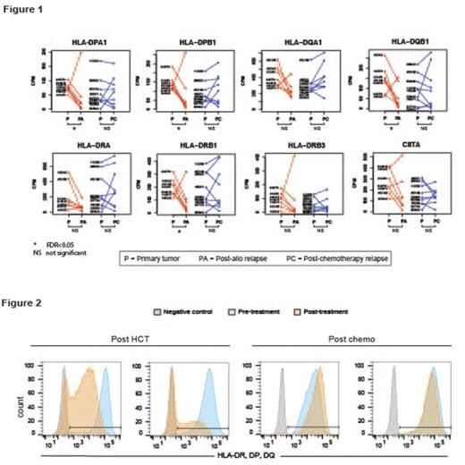
Four classical MHC Class II genes were among the 187 genes that were downregulated after post-HCT relapse: HLA-DPA1 (mean fold change -11.9 in 6/7 cases), HLA-DPB1 (mean fold change -6.5 in 6/7 cases), HLA-DQB1 (mean fold change -9.7 in 6/7 cases), and HLA-DRB1 (mean fold change -11.9 in 6/7 cases) (Figure 1). In these same 6 cases, there was a strong (but not significant) trend towards downregulation in the remaining MHC Class II genes: HLA-DQA1, HLA-DRB3, and HLA-DRA, as well as CIITA, a transcriptional activator believed to act as the "master regulator" of MHC Class II genes (Figure 1). Several other genes involved in MHC Class II antigen processing and presentation were significantly downregulated post-HCT (FDR<.05), including HLA-DMA, HLA-DMB, and CD74 . In contrast, there was no significant downregulation of MHC Class I genes observed after allo-relapse, raising the possibility that loss of MHC Class II presentation may play a particularly important role in immune evasion after HCT. To validate these findings, we performed flow cytometry for Class II and Class I genes on banked post-HCT relapse samples, and this confirmed significant downregulation of MHC Class II (but not Class I) expression on post-HCT relapse samples (Figure 2). Taken together, these results suggest that loss of MHC Class II-mediated antigen presentation may be an important mechanism of immune escape after HCT.
No relevant conflicts of interest to declare.
Author notes
Asterisk with author names denotes non-ASH members.


This feature is available to Subscribers Only
Sign In or Create an Account Close Modal